breast cancer
breast mass icd 9 breast mass biopsy breast massage therapy breast massage growth breast mass in male breast mass excision breast mass removal breast mass size breast mass ultrasound breast mass with pain
Sunday, May 24, 2015
Sunday, May 17, 2015
The breast:
is an apocrine gland containing the mammary gland that produces milk to feed an infant child.•Both men and women develop breasts from the same embryological tissues.
•However, at puberty, female sex hormones, mainly estrogen, promote breast development, which does not occur in men, due to the higher amount of testosterone.
• the breast is a cone with the base at the chest wall, and the apex at the nipple.
• nipple: is a small projection
of skin containing the outlets for 15–20 lactiferous
ducts arranged cylindrically around the tip.
• Areola: the pigmented area on the
human breast around the nipple.
• Each breast contains 15–20 lobes. The subcutaneous adipose tissue covering the lobes gives the breast its size and shape.
• Each lobe is composed
of many lobules, at the end of which are sacs where milk is produced in
response to hormonal signals.
• the
weight of the breast 500–1,000 grams.
•Sensation
in the breast is provided by the peripheral nervous system innervation, by cutaneous branches of the
fourth-, the fifth-, and the sixth intercostal nerves.
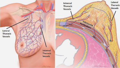
• vascularisation:
• Lymphatic drainage: 75% of
the lymph from the breast travels to the ipsilateral axillary lymph nodes,
whilst 25% of the lymph travels to the parasternal nodes.
Types of Breast Lumps
A)-
Cysts and Abscess Lumps:
• breast cyst is a non-cancerous, fluid-filled sac in the breast. They generally feel smooth or rubbery under the skin and can be quite painful or cause no pain at all. Cysts are caused by the hormones that control the menstrual cycle and are rare in women older than 50.
• sebaceous cyst is a non-cancerous, closed sac or cyst below the skin that is caused by plugged ducts at the site of a hair follicle. Hormone stimulation or injury may cause them to enlarge but if no symptoms are present, medical treatment is not required.
• Abscesses are non-cancerous pockets of infection within the breast. They can be quite painful and cause the skin over the breast to turn red or feel hot or solid. Abscesses of the breast are most common in women who are breast-feeding.
B)- Growths:
• Adenomas are non-cancerous abnormal growths of the glandular tissue in the breast. The most common form of these growths, fibroadenomas, occur most frequently in women between the ages of 15 and 30 and in women of African descent. They usually feel round and firm and have smooth borders. Adenomas are not related to breast cancer.
• Intraductal papillomas are wart-like growths in the ducts of the breast. These lumps are usually felt just under the nipple and can cause a bloody discharge from the nipple. Women close to menopause may have only one growth, while younger women are more likely to have multiple growths in one or both breasts.
• Breast cancer usually feels like a hard or firm lump that is generally irregular in shape and may feel like it is attached to skin or tissue deep inside the breast. Breast cancer is rarely painful and can occur anywhere in the breast or nipple
C)- Fatty Lumps:
• Fat necrosis is a condition in which the normal fat cells of the breast become round lumps. Symptoms can include pain, firmness, redness, and/or bruising. Fat necrosis usually goes away without treatment but can form permanent scar tissue that may show up as an abnormality on a mammogram.
• lipoma is a non-cancerous lump of fatty tissue that is soft to the touch, usually movable, and is generally painless.
-:Caution :-
- When women feel a lump 36 of 100 are real
- Women with true lump
- Not dangerous or “benign” 85 out of 100
- Cancer 15 out of 100
Evaluation of breast mass
History:- Change in general appearance of breast (size, symmetry)
- New or persistent skin changes
- New nipple inversion
- Breast pain (cyclic vs. noncyclic, duration, location in breast)
- Breast mass (how it was discovered, duration, change in size, location)
- Relationship of mass to menstrual cycles
- Nipple discharge (unilateral vs. bilateral, color)
- Medications (e.g. hormones)
- Risk factors for breast cancer
Clinic exam
Inspect (relaxed, arms raised, hands on hips)
Breast symmetry
Skin changes (dimpling, retraction, edema, ulceration)
Nipples (symmetry, inversion/retraction, discharge)
Palapation (breasts, axillae, entire chest wall)
Pain
Masses
Regional lymph nodes (Axillary and Supraclavicular)
Workup
•Imaging
•Bilateral
diagnostic mammogram (>30)
•U/S
– solid vs cystic (<30)
•Biopsy
–Palpable
– FNA or CNB
–Non-palpable
- stereotactic or ultrasound-guided percutaneous core biopsy
–Incisional biopsy
Nipple discharge
Etiology
Lactation
Physiologic nipple
discharge
- Hyperprolactinemia
- Hypothyroidism
- Medication related
- Neurogenic stimulation
Pathologic
- Intraductal papilloma
- Ductal ectasia (common cause)
- DCIS
•Investigation:
1-cytology of discharge for exfoliated cancer cells.
2-mammography
3-duct galactography
4- serum prolactine
•Treatment:
1-palpable mass ==== excised for histology and treated
accordingly.
2-single duct bloody discharge === microdochectomy
3-multiple ducts no mass ==initially conservative by
observation.
If persiste === excision of
major ducts
Breast cancer
Risk Factors vs.
Protective Factors
Neoplasia and benign Tm
Duct
papilloma
•Usually in the main
ducts near the nipple.
•Young women.
•Before it become
palpable may ulcerate and bloody discharge.
•Block may cause a
retention cyst.
Clinic :
1- bloody
discharge (no pain).
2- small mass fusiform behind areola. Pressur on it bloody
discharge.
3- if no mass the
pressure on certain point behind the areola will reveal discharge from one duct
•Investigation: Ductography
•Treatment: Excision of the
affected duct (micro-dochectomy).
fibroadenoma
The commonest breast mass of young women (15-30)
Fibrous +++ and glandular tissues ++
clinical:
Hard mass (20-30) painless , soft mass (30-50) painfull , accident discover
•Usually
small, non tender, spherical, firm, well circumscribed, high mobility.
•Investigation:
Clinical exam enough for diagnosis
Soft tissue == mammography ==reveal a well
circumscribed.
May U/S
Treatment: Excision and
histological confirmation of diagnosis
CYSTOSACROMA phylloides
-A highly cellular type of fibroadenoma that tend to grow
rapidly (brodie). Enlarge slowly and
rich a large size (20-30 cm).
-Cystic formation with skin ulceration
-Rarely malignant.
-Phylloides tumour
-wide local
excision to prevent recurrence
-if infiltrate whole breast simple mastectomy.
Carcinoma of breast
•Most
common cancer in women
•In
USA 9:1
•Breast
cancer is second only to lung cancer as a cause of cancer deaths in American
women
•The
mean age of affection 60 years.
•The
commonest malignant neoplasm in Egyptian female 35%.
Histology:
•The
carcinoma may arise from the lobule, ducts or nipples
•Arise
from the ductal epithelium in the majority of cases.
Carcinoma of
ducts:
-ductal carcinoma in situ
-Infiltrating ductal carcinoma
•Carcinoma of the
lobules:
-Non infiltrating
lobular carcinoma (lobular carcinoma in situ)
-Infiltrating lobular
carcinoma. Bilateral in 25%.
•Paget’s
disease of nipple:
An introduction of carcinoma which begins in the
epithelium of a main collecting lactiferous duct and
spreads within epithelium
up to the skin of the nipple and down into the breast substance.
A mass may appear after 2 years from the start of the
disease.
•Spread:
Local
spread: overlying (skin),
underlying (pectoralis major,…..., chest
wall).
Lymphatic
spread: mostly to axillary node, next common is
the internal mammary chain.
Blood
stream spread: produces
metastases in the lungs, bones, brain and liver.
Clinical features
•Symptoms:
1- accidently
notices a painless lump.
2- mild breast
pricking pain , nipple retraction or bloody nipple discharge.
3- symptomatic
metastases.(axillary lump ,pulmonary
metastasis)
4- by routine
screening mammography in high risk women.
•Signs:
For breast examination , the top half of trunk exposed,
both breast, axillae, arms and supraclavicular.
-Beast asymmetry - enlargement
-Skin dimpling - skin puckering
-Peau d’orange - skin ulceration or
nodule
-Mass : hard ,
irregular , immobile
, fixe
Nipple : retraction ulce
STAGING OF cancer breast
•Manchester
staging:
Stage 1:
Mobile mass , no LNs , no
attachment of wall.
Stage 2:
Mobile mass , no attachment of wall , mobile ipsilateral axillary LNs.
Stage 3: (wide licale spread) any of theses
1-skin affection (more than tm but at breast)
2-fixed to pectoral muscles
3-ipsilateral axillary LN
matted together
4-ipsilateral supra clavicular LNs
5-edema of the arm.
Stage 4:
1-skin affection wide of the breast.
2-fixed to chest wall.
3-controlateral axillary LNs.
4-distant metastasis.
v tmn:
Subscribe to:
Posts (Atom)


